Diagnostics
consulting and examination
Open hours:
- CT/MRI: 09:00 a.m. - 06:00 p.m.
- X-Ray: 09:00 a.m. - 03:00 p.m.
- Other departments: 09:00 a.m. - 04:00 p.m.
- On holidays – upon agreement.
Address:
- 27 Bulvarno-Kudriavska Street,
Kyiv 01054 Ukraine - 7 Chekhivsky Lane,
Kyiv 01054 Ukraine.
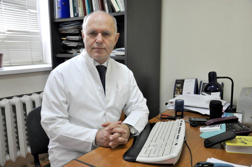
 Chief —
Haiko Oksana Dr.hab.med
Staff of the department consists of 6 scientific researches, among them 2 doctors of sciences, 4 candidates and 6 doctors.
The premises of the Department are the basis for the Centre of Osteoporosis, providing diagnostic, prevention and treatment to adults and children with diseases accompanied by osteoporosis.
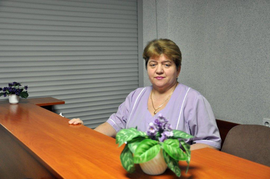
Staff members of the department perform examinations and consult patients on daily basis.
Each year over 10 000 underwent examination by the abovementioned technologies, and over 15 000 studies are made.
The department participates actively in scientific and research work of the Institute. It published over 400 scientific works, inter alia 3 monographs. Priority directions of the department’s work is early diagnostics of congenital pathologies of hip joint, traumatic injuries of joints and peri-articular structures, diagnostics and treatment of nerve and muscle pathology in patients with different orthopedic diseases and trauma injuries of skeleton.
The 3rd East-European Congress of pain
Main directions of scientific-practical work:
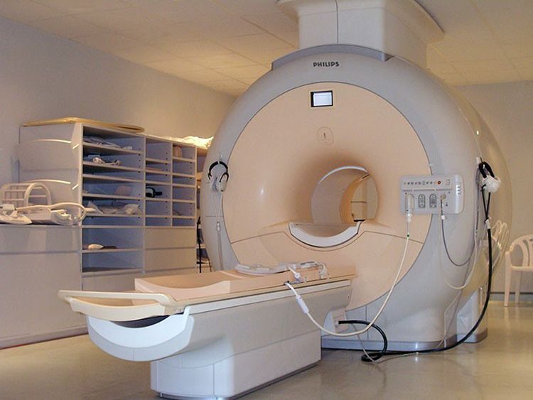
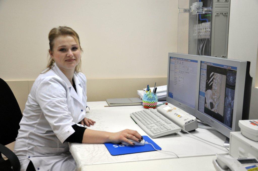
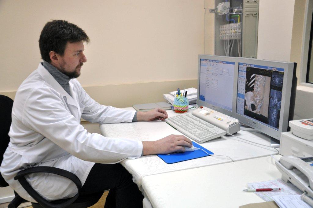
MRI
In our department, the magnetic resonance imaging is carried out on the Philips Achieva 1.5 Tesla, channel, (production in the Netherlands). This machine combines everything you need to get a high quality image.

CT
CT is fast, secure and painless method to receive complete information about a human body. Results of CT examination can be handed to a patient at the same day.
X-Ray
X-ray examination is the method of non-invasive radiological studies of anatomic structures of a body.
X-Ray densitometry
Double energy X-ray absortiometics (DXA). It is the “golden standard” for diagnosis of osteoporosis.
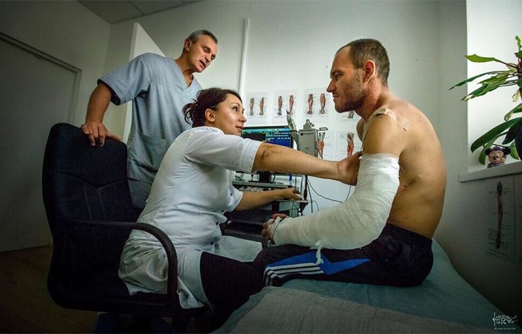
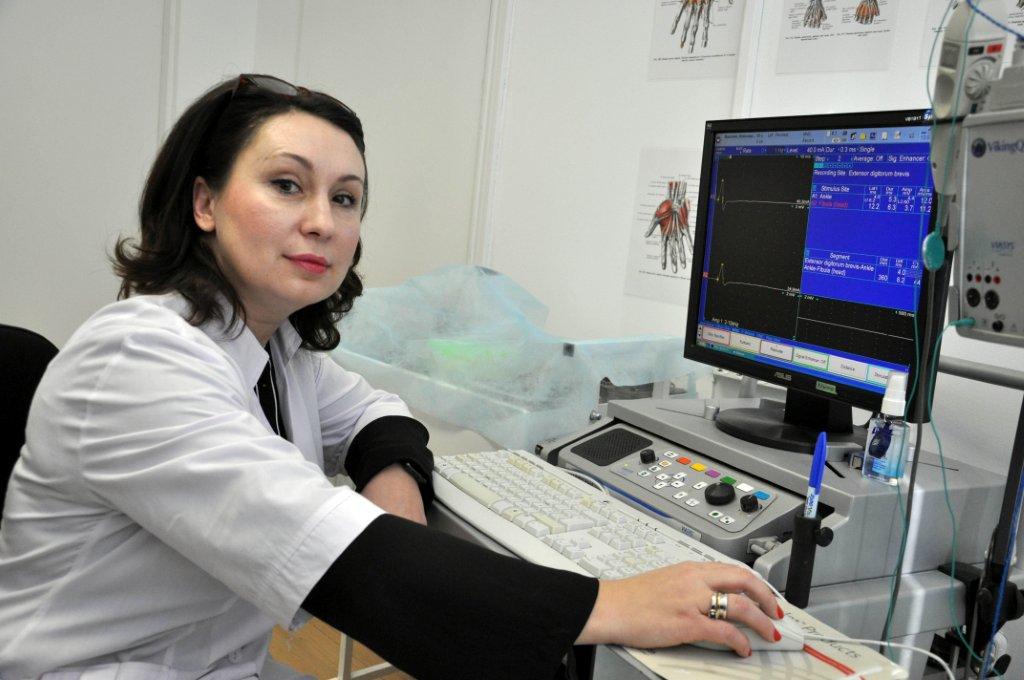
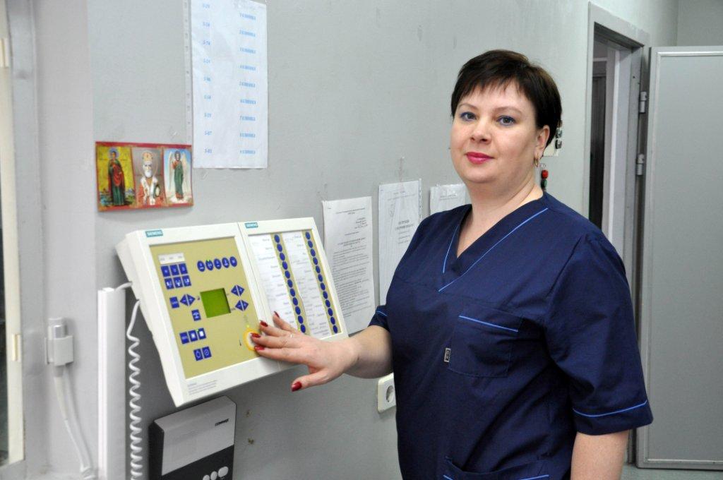
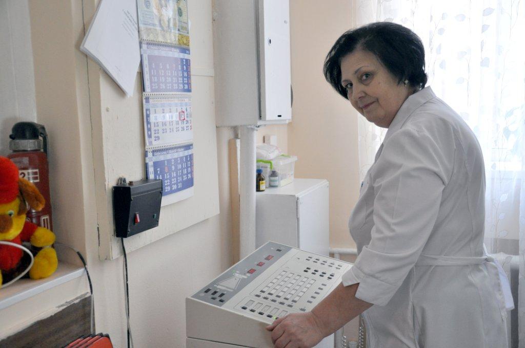
Electromyography
EMG is a diagnostic procedure, intended to estimate conditions of muscles, nerves and nerve cells that control thereof. The method helps to define the grounds of frequent problems lime muscle weakness of extremities, disorders of sensitivity (sleep etc.)
Reovasography
Reovasography is the method of study, reflecting condition of general regional blood flow in studied organ, based on registration of fluctuation of resistance in a live tissue of a body to high frequency current, registration of pulse fluctuations of vessels. Reovasography is applied to control the efficiency of treatment as well as for diagnostics.
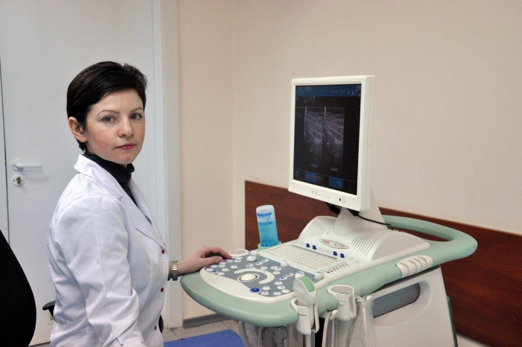
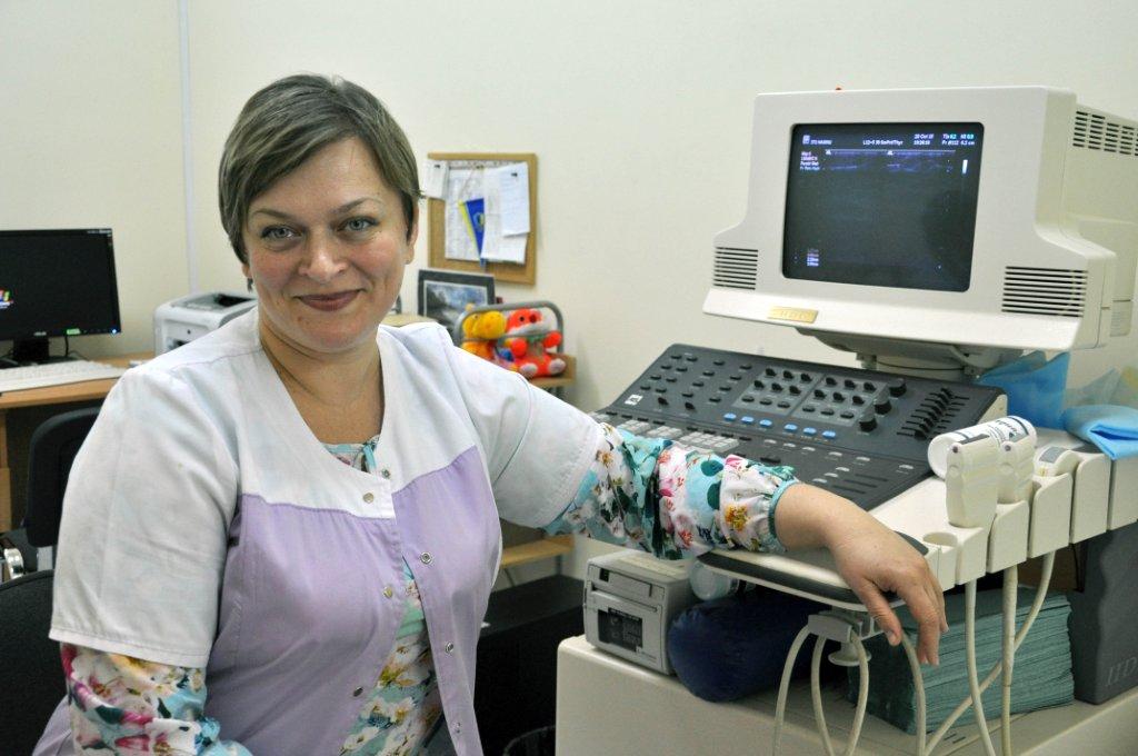
Doppler sonographic
The modern examination method for blood vessel examination, an ultrasound dopplerography (USDG) gives a possibility do define conditions of your vessels and to assess the risk of vessel system diseases development.
US
Diagnostics of locomotion system disorders in children under 1 y.o., US of abdominal cavity organs and retroperitoneal space, US of locomotion system
Laboratories
microbiology and chemotherapy, neuro-orthopedics and problems of pain, biochemistry, biomechanics, immunology and Pathomorphology department with biologic subdivision (vivarium)


- Tag:
- MRI
- magnetic resonance imaging
- Computer tomography services
- Computer tomography
- X-Ray
- X-Ray densitometry
- Electromyography
- Reovasography
- Electropuncture diagnostics
- Electropuncture diagnostics by R.Foll
- Doppler sonographic
- ultrasound diagnostics
- US
- US of locomotion system
- Diagnostics of locomotion system disorders
- US of abdominal cavity organs
- US retroperitoneal space
- laboratory
- diagnostics of joints
- diagnostics of fractures
- diagnostics of traumatic injuries