Нallux valgus
Нallux valgus deformity (Нallux valgus) is a deforming disease of the first toe, which is characterized by a progressive course, which changes its position relative to the anatomical axis of the foot and other toes, the language of the average citizen "bump on the feet" - is one of the foot deformities
Published: 11.09.2020
Нallux valgus deformity
Hallux valgus is a deforming disease of the first toe, which is characterized by a progressive course, which changes its position relative to the anatomical axis of the foot and other toes, the language of the average citizen "bump on the feet" - is one of the foot deformities. Valgus deformity leads to inflammation in the area of the joint bursa, with exacerbation of pain on the inner surface of the first toe. Deformation of the foot’s front parts leads to impaired walking. It is most common among women.
The cause of valgus deformity of the first toe is:
- Connective tissue weakness;
- Hormonal damage;
- Genetic skill;
- Long-term wearing of tight model shoes;
- Shoes with high heels and a narrow toe;
- Wrongly chosen shoes.
Treatment
The most effective method of treatment is a surgery. A number of operative techniques are involved: on soft tissues (McBride's operation) and on bones (SCARF, Chevron, Akin, Hohmann, Kramer, Mau, Wilson, proximal osteotomy), arthrodesis of the foot’s bones and combined injuries.
Postoperative period
In the postoperative period, the patient is activated the day after surgery in special Baruk’s shoes. It is necessary to spend from 4 to 8 weeks in orthopedic shoes, depending on the performed surgical methods. Treatment in the hospital - 3-4 days. Suture removal after 14 days. It is not desirable to wear shoes with heels larger than 3-4 cm up to 6 months. The period of stay on the sick-leave is 1.5-2 months.
Clinical case
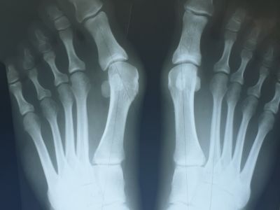
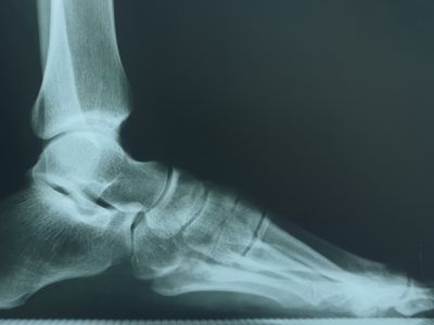
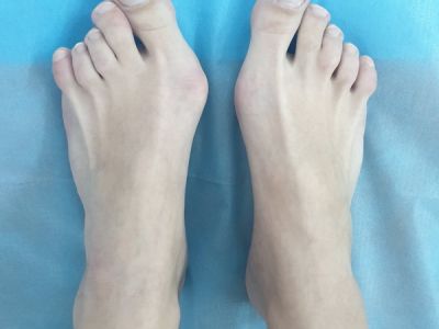
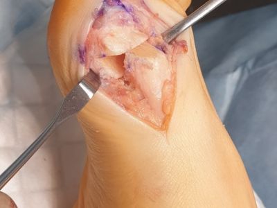
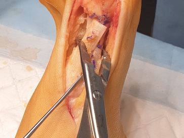
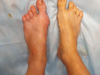
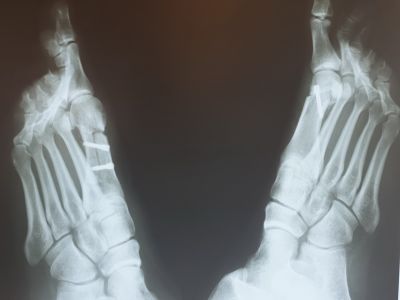
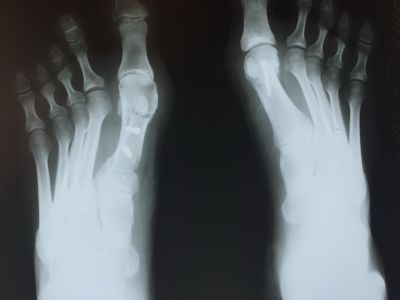
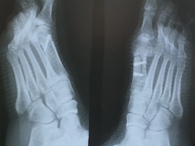
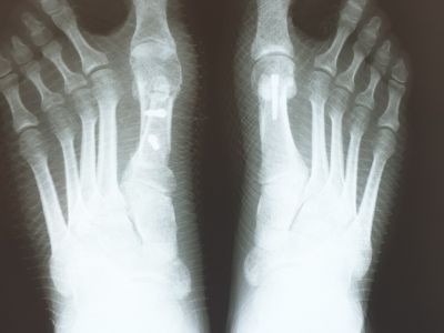
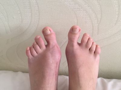
The patient consulted with complaints of foot pain after exercise and walking in high heels. Surgery was performed after clinical and instrumental examination: SCARF osteotomy on the left foot and Chevron osteotomy on the right foot. The figure shows photos and X-ray before surgery, during the operation and the appearance of the feet 1 year after surgery.
